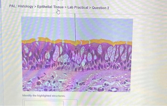
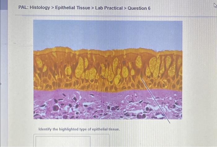
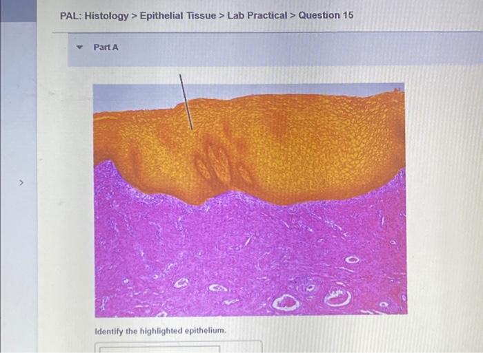
Embarking on pal histology epithelial tissue lab practical question 1, this discourse delves into the intricate realm of oral histology, meticulously examining the histological features, laboratory techniques, and clinical applications of palatal epithelial tissue. Brace yourself for an enlightening journey that unravels the complexities of this specialized tissue.
Palatal epithelial tissue, a stratified squamous epithelium lining the palate, plays a crucial role in oral health and function. Its histological features, including its distinct layers, cell types, and specialized structures, provide valuable insights into its physiological functions. Laboratory techniques, such as tissue preparation, staining methods, and microscopy, empower researchers to study this tissue in detail, enabling a deeper understanding of its structure and function.
Histological Features of Palatal Epithelial Tissue: Pal Histology Epithelial Tissue Lab Practical Question 1

The palatal epithelium is a stratified squamous epithelium that lines the hard and soft palate. It is composed of several layers of cells, including the basal layer, spinous layer, granular layer, and cornified layer. The basal layer is the deepest layer and is composed of cuboidal or columnar cells that are attached to the basement membrane.
The spinous layer is located above the basal layer and is composed of polygonal cells that are connected by desmosomes. The granular layer is located above the spinous layer and is composed of cells that contain keratohyalin granules. The cornified layer is the outermost layer and is composed of dead cells that are filled with keratin.The
palatal epithelium has a number of functions, including protection, lubrication, and sensation. The basal layer of the epithelium is responsible for cell renewal, while the spinous layer provides strength and flexibility. The granular layer helps to protect the epithelium from dehydration, while the cornified layer provides a waterproof barrier.
The palatal epithelium also contains a number of sensory receptors that are responsible for taste and touch.
Laboratory Techniques for Studying Palatal Epithelial Tissue
There are a number of laboratory techniques that can be used to study palatal epithelial tissue. These techniques include tissue preparation, staining methods, and microscopy.Tissue preparation involves removing the palatal epithelium from the underlying tissue and then fixing it in a preservative solution.
The most common fixative used for palatal epithelial tissue is formalin. Once the tissue has been fixed, it can be embedded in paraffin or frozen for sectioning.Staining methods are used to visualize the different components of the palatal epithelium. The most common staining method used for palatal epithelial tissue is hematoxylin and eosin (H&E).
H&E staining stains the nuclei of the cells blue and the cytoplasm pink. This allows the different layers of the epithelium to be easily distinguished.Microscopy is used to examine the stained tissue sections. The most common type of microscope used for palatal epithelial tissue is the light microscope.
Light microscopes use visible light to illuminate the tissue sections. This allows the different components of the epithelium to be seen in detail.
Practical Question 1: Histological Analysis of Palatal Epithelial Tissue
Specimen:Palatal epithelium from a human cadaver Staining Method:Hematoxylin and eosin (H&E) Analysis Parameters:* Identify the different layers of the palatal epithelium
- Describe the cell types found in each layer
- Discuss the functions of the different layers of the palatal epithelium
Clinical Applications of Palatal Epithelial Tissue Analysis, Pal histology epithelial tissue lab practical question 1
Palatal epithelial tissue analysis is used in a variety of clinical applications, including the diagnosis and management of oral diseases. For example, palatal epithelial tissue biopsy can be used to diagnose oral cancer, oral lichen planus, and other oral diseases.
Palatal epithelial tissue analysis can also be used to monitor the response to treatment for oral diseases.
Research Frontiers in Palatal Epithelial Tissue Biology
There are a number of research frontiers in palatal epithelial tissue biology, including stem cell research, tissue engineering, and regenerative medicine. Stem cell research is focused on the development of new methods to grow and differentiate palatal epithelial cells. Tissue engineering is focused on the development of new methods to create artificial palatal epithelial tissue.
Regenerative medicine is focused on the development of new methods to repair and regenerate damaged palatal epithelial tissue.
FAQ
What is the significance of palatal epithelial tissue?
Palatal epithelial tissue serves as a protective barrier against mechanical, chemical, and microbial insults, ensuring the integrity of the oral cavity.
How does histological analysis contribute to our understanding of palatal epithelial tissue?
Histological analysis allows us to visualize the microscopic structure of palatal epithelial tissue, revealing its cellular composition, tissue architecture, and specialized features.
What are the key laboratory techniques used to study palatal epithelial tissue?
Tissue preparation, staining methods (e.g., hematoxylin and eosin), and microscopy are essential techniques for examining the histological features of palatal epithelial tissue.


